baby chest x ray technique
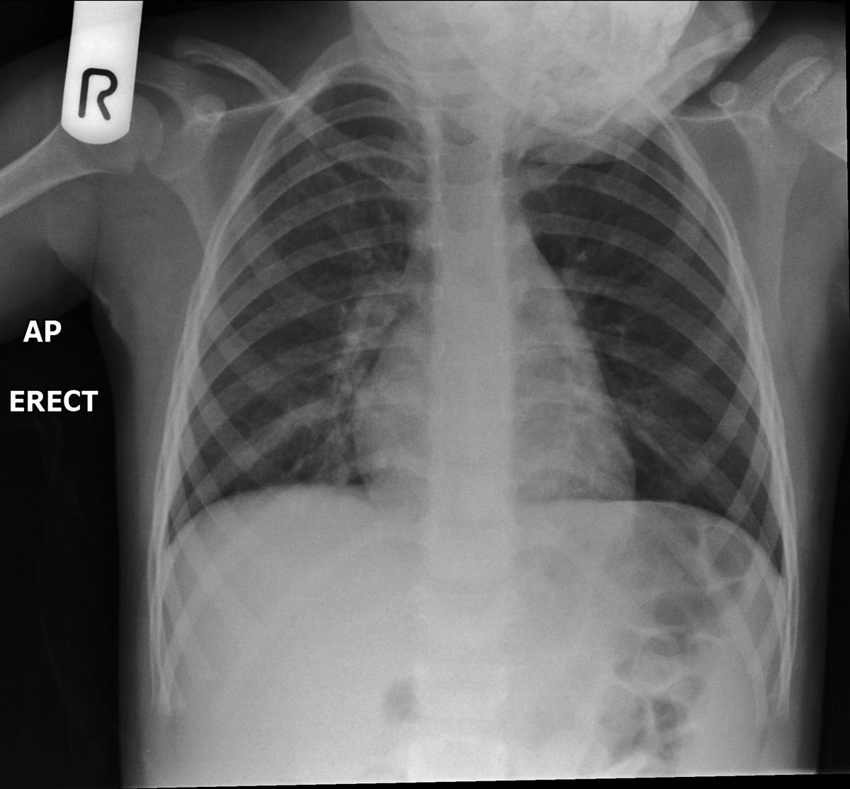
The publication of this study and exposure chart could act. Up to 10 cash back The radiographer inside the cubicle positions the bed close to the glass door and places the digital detector behind the patient for an erect anteriorposterior chest X-ray before stepping away from the patient during the exposure a while the radiographer outside the cubicle positions the X-ray tube head close to the glass door and steps laterally.

Ce4rt Guide For X Ray Techs To Immobilize Pediatrict Patients
The Chest Radiograph.

. Frontal chest radiographs are widely performed. A chest radiograph for a 12-year-old female is an embarrassing ordeal. Chest PAAP erect 180 cm.
The aim of this study was to develop and validate a prediction model to estimate the probability for a normal chest x-ray in children with RSV infection. Erect chest X-rays are taken at 180 cm. Hospi-tals were excluded if they did not have an x-ray department a mobile x-ray machine or refused participation.
In Canada 33 hospitals were contacted initially as potential participants. 901a a Use automatic exposure control 500 speed for chestabdomen else 400 speed at specified kVp when practical. However all children are modest to some degree about having their genitals or backsides exposed after ages 4 to 5.
The plain chest radiograph remains the first radiological examination in use for the evaluation of the chest in children. Mearadji International Foundation for. 851a a Use automatic exposure control 500 speed for chestabdomen else 400 speed at specified kVp when practical.
In contrast most 12-year-old males have little modesty about their chests. Chest AP erect in chair 180 cm. Everytime I have taken him to the Drs they have said its a virus hes struggling to shake off.
A chest x-ray is frequently performed in infants with LRTI caused by RSV. Full legfull spine imaging is performed at 180 cm using CR. In most cases rib X-rays are performed in frontal and lateral projections.
The normal neonatal chest X-ray. If we are obviously talking about any part of the chest then a targeted X-ray of the affected ribs is performed. As a benchmark for other medical imaging departments and to promote discussion on digital X.
When I took him back this morning we saw a different Dr and she listened to his chest and said it sounded clear but. Patient Position Infant. GE AMX4 portable x-ray system Fuji CR imaging plates and reader Tracked AP chest and abdomen for patients 0-3 months in the NICU and PICU at Hadassah Medical Organization Image quality assessment and dose estimation for high and low kVp image sets.
Pediatric Chest Screen 70-80 DIGITAL OPTIMUM kVp Universal CR Technique Chart using a standard 21 LgM Part View kV mAs kV mAs kV mAs Abdomen AP Grid 85 10 -15 85 20 - 25 85 30 - 40 Ankle AP 70 18 70 2 70 25 Ankle Obl 70 16 70 18 70 22 Ankle Lat 70 15 70 16 70 2 Chest -Adult AP 400 - tt -72 85 2 - 25 85 32 - 4 90 5 - 64. Lateral cervical spines are taken at 150 cm. Chest radiographs Systematic review of chest radiographs is necessary for accurate evaluation.
12 important topics 1. The target population of this study is imaging de-partments representing all geographic regions in Canada and Norway that perform mobile chest radiographs. Use gonadal shielding if possible.
Immobilize infants arm above the head or use stockinette ace bandage tape or sandbags. Center Image receptor to Central Ray. This approach allows you to assess the overall condition of the breast.
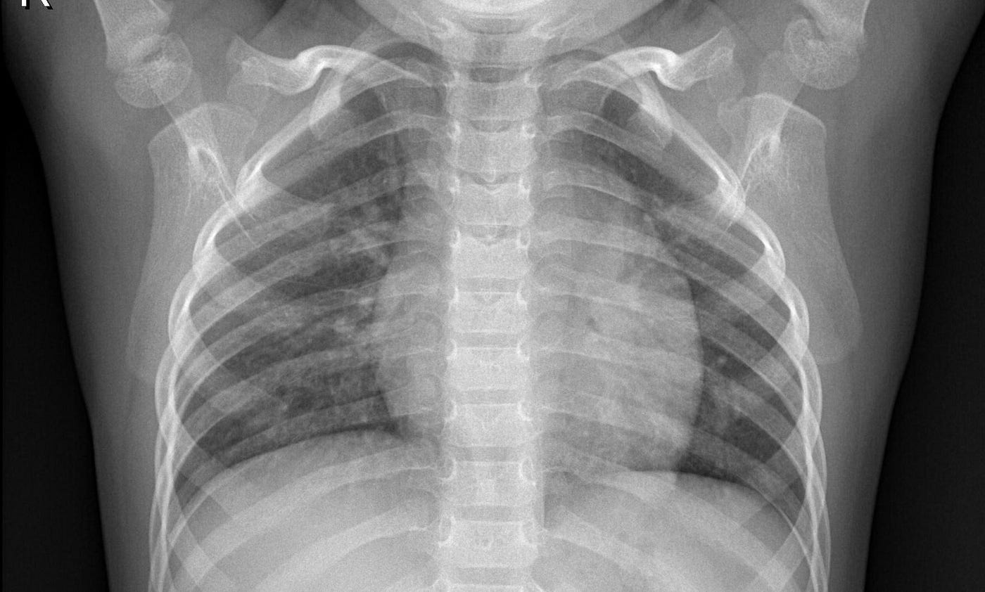
Most neonatal chest X-rays are AP films unless the baby is made to lie prone Lucency of soft tissue shadow - darker the soft tissue more is the exposure Ease of visibility of retrocardiac vertebrae if the retrocardiac vertebrae are easily seen the film is over exposed Relative lucency of lung fields. The American Dental Association ADA recommends that kids and teens get bitewing X-rays every six to 12 months if they have cavities. This is partly due to modesty but also due to fear.
Radiologists consider a chest X-ray to be of good quality when the trachea is centered and equidistant from the head of the clavicle on both sides the spine is visible as a transparent structure through the heart shadow and there is full inspiratory effort the right 6th rib is at the midpoint of the hemidiaphragm on that side. Baby with persistant cough - Dr has suggested a chest x-ray. Pediatric chest X-ray M.
My 8 month old baby has had a persistant cough since he was 612 months. Aerationofthenormalneonatallungisvirtuallycomplete within two or three respiratory cycles after birth and the lung fields should appear symmetrically aerated on the initial X-ray with the diaphragms lying at the level of. Immobilize legs with Ace bandage or tape and sandbags.
Lateral views tend only to be performed after review of the frontal radiograph when there are unanswered clinical questions. All distal extremity exposures are taken at 110115 cm SID.
X Ray Image Classification The Easy Way By Amanda Woo Towards Data Science

Neonatal Chest Radiograph In The Exam Setting Radiology Reference Article Radiopaedia Org

Pediatric Chest Horizontal Beam Lateral View Radiology Reference Article Radiopaedia Org

T Spine Image Radiologic Technology Radiology Tech Radiology Technologist

Diagnosis Of Other Lung Conditions In Premature Babies

Pediatric Chest Supine View Radiology Reference Article Radiopaedia Org
Paediatric Chest Immobilisation Devices Wikiradiography

Chest X Ray Of A 6 Month Old Child With An Icd The Active Can Is Download Scientific Diagram
2

Diagnosis Of Other Lung Conditions In Premature Babies

The Normal Cxr Nurse Radiology Nursing Education
2

An Ap Erect Chest Xray Of The Patient Showing An Upward Tilted Cardiac Download Scientific Diagram

Pin By Eve Emmanuelle Roy Hebert On T I M Diagnostic Imaging Med Student Science Degree
Chest X Ray Of The Neonate On First Day Of Admission Showing Multiple Download Scientific Diagram

Osteopoikilosis Spotted Bone Disease Radiology Imaging Nuclear Medicine Technology Bone Diseases

Chest X Ray Appearances In Ventilated Infants With Mas A Typical Download Scientific Diagram

Medial Segment Of Right Middle Lobe Consolidation Radiology Case Radiopaedia Org Radiology Imaging Radiology Segmentation

How To Read Chest X Rays International Emergency Medicine Education Project